
דף הבית » Melanoma and Skin Cancer

Skin cancer refers to a group of diseases in which skin cells grow uncontrollably and form malignant tumors
It is one of the most common types of cancer, but in most cases it can be successfully treated if diagnosed early. Common skin cancers can be broadly divided into two main types: melanoma, and non-melanoma skin cancers, primarily basal cell carcinoma (BCC) and squamous cell carcinoma (SCC).
Melanoma is a type of cancer that usually begins in the skin. More specifically, it arises from pigment-producing cells called melanocytes — the same cells that give our skin its color and form benign moles. Melanoma typically appears as a brown or black skin lesion that starts flat and becomes raised over time. These lesions can be multicolored (black, brown, gray, blue, red, white) or, less commonly, pink or skin-toned. They are usually asymmetrical and grow faster than other skin lesions.
Melanoma usually begins with a change on the skin — either a new mole or a change in an existing one. In such cases, you should see a dermatologist promptly. Warning signs in existing moles include:
• Color – A lesion with multiple colors (brown, black, gray, red, white).
• Asymmetry – When one half of the lesion is different from the other.
• Border – Irregular or poorly defined borders.
• Diameter – Moles larger than 5 mm.
• Evolution – Lesions that grow or change in color, shape, or characteristics over weeks or months.
• Ugly duckling – A lesion that clearly stands out from other skin lesions.
Early detection saves lives — melanoma is often curable when diagnosed early!
Each year, around 2,000 new cases of melanoma are diagnosed in Israel. Approximately 2–3% of the Jewish population in Israel is expected to develop melanoma in their lifetime.
Early-stage melanoma has excellent survival rates — 98% five-year survival. In contrast, late-stage melanoma has a five-year survival rate of only about 30%. Early diagnosis and appropriate treatment are the keys to recovery and survival.
Melanoma arises when DNA mutations accumulate in melanocytes, disrupting their division control and turning them cancerous. A major cause of cutaneous (and ocular) melanoma is ultraviolet (UV) radiation from sun exposure or tanning beds. Repeated UV exposure, especially with sunburns, is the most significant risk factor. However, melanoma can also develop without sun exposure.
Risk factors include:
• Personal history of melanoma.
• Family history (first-degree relatives) of melanoma.
• Multiple moles.
• Atypical (dysplastic) moles.
• History of non-melanoma skin cancers like BCC or SCC.
• Severe sun-damaged skin.
• Genetic syndromes (e.g., BRCA2 and others).
• Immunosuppression due to illness or immunosuppressive medications (e.g., autoimmune diseases, organ transplants).
Other risk factors: fair skin, red hair, blue or green eyes, and history of sunburns. Even if you’re not in a high-risk group, stay alert to new or changing lesions.
Superficial Spreading Melanoma – The most common type; typically starts as a flat, slow-growing lesion.
• Nodular Melanoma – Faster-growing, often appears on head, neck, chest, or back.
• Lentigo Maligna Melanoma – Grows slowly over years, often evolving from a precancerous lesion on sun-exposed areas like the face or neck in older adults.
• Acral Melanoma – Rare, appears on palms, soles, or as a dark streak under nails; more common in darker skin tones, not sun-related, and more aggressive.
• Mucosal Melanoma – Rare; appears in internal mucosal surfaces (e.g., nose, mouth, GI tract, rectum).
• Uveal Melanoma – Arises within the eye; behaves differently from cutaneous melanomas.
The combination of early detection and advanced drug therapies provides many reasons for optimism in patients at increased risk for melanoma. The recommended treatment approach depends on the stage of the disease, making accurate staging essential. Our expert team determines the optimal treatment plan in collaboration with the patient.
Staging is essential to understand the risk posed by the disease and to plan appropriate treatment and follow-up. It is based on pathology report findings and assessment of lymph node and organ involvement. The oncologist leads this critical process.
Pathology features include tumor depth (in millimeters), presence or absence of ulceration, and mitotic rate. These help define the tumor’s aggressiveness. The oncologist will explain these findings in detail.
Internal organ involvement is assessed with imaging tests like PET-CT and brain MRI. PET-CT reveals abnormal metabolic activity suggestive of metastasis. Brain MRI identifies metastases in the brain with high precision. Imaging recommendations are tailored to the patient and tumor features.
The lymphatic system functions as the body’s drainage and immune surveillance system. Melanoma cells can spread through it. Evaluation includes imaging and possibly sentinel lymph node biopsy (SLNB) to determine if the lymph nodes are affected.
What is a sentinel lymph node biopsy (SLNB)?
Even when lymph nodes appear normal on imaging, they may contain microscopic melanoma cells. The likelihood of this increases with tumor depth — roughly 10% per 1 mm of invasion. SLNB is usually recommended when invasion is greater than 0.8 mm, though exceptions may apply.
The sentinel node is the first lymph node draining the tumor site. It is identified using a dual-tracer method: a small radioactive substance and blue dye are injected near the melanoma site, and the surgeon identifies and removes the blue node during surgery. This node is then examined microscopically.
If cancer cells are absent, other nodes are unlikely to be involved. If present, it confirms early spread and helps guide further treatment.
Melanoma is potentially lethal, but with early diagnosis and modern therapies, patients have many reasons to be hopeful. Early-stage melanoma often has little to no impact on quality or length of life. More advanced cases may require combined treatment approaches.
How is thin localized melanoma (up to Stage IIa) treated?
Melanoma in situ is excised with 5 mm margins. Tumors less than 0.8 mm deep (Stage IA) are excised with 1 cm margins. Sentinel node biopsy or imaging may be discussed. Deeper tumors often require SLNB. Surgery is usually sufficient, with no need for further therapy.
How is thick localized melanoma (Stage IIb–IIc) treated?
These cases require excision with 2 cm margins and SLNB. Even if nodes are clear, adjuvant immunotherapy is recommended to lower the risk of recurrence. The oncologist tailors the treatment plan.
How is regional melanoma (Stage III) treated?
Lymph node involvement indicates high metastatic potential. If detected microscopically (positive SLNB), immunotherapy or targeted therapy is given post-surgery. If detected by imaging or clinical exam, surgery plus drug therapy is needed. Radiation is generally not required.
How is metastatic melanoma (Stage IV) treated?
Although the most serious stage, it can be managed effectively. Treatments include immunotherapy, targeted therapy, and occasionally focused radiation.
What is immunotherapy for melanoma?
הטיפולים אפקטיביים ומביאים לעיתים קרובות לנסיגת מחלה הנמשכת שנים רבות ואף להביא לריפוי. התאמת הטיפול והליווי נעשים ע”י האונקולוג בהתאם למאפייני המחלה והמטופל. ניתן להשתמש באימונותרפיה גם כטיפול מניעתי לאחר ניתוח וגם כטיפול במחלה גרורתית.
Approximately 50% of melanomas have a BRAF mutation. Targeted drugs block the BRAF and MEK proteins to stop tumor growth. Oral combinations include Braftovi + Mektovi, Tafinlar + Mekinist, and Zelboraf + Cotelic. These treatments are fast-acting and generally well-tolerated. They may be used after surgery or for metastatic disease, with plans guided by the oncologist and sometimes a dermatologist.
In advanced cases or if standard therapies fail, off-label or personalized treatments may be considered. Expertise in melanoma biology and access to innovative oncology options can open new doors.
Patients are at dual risk:
First- and second-degree relatives are also at increased risk. They should undergo regular dermatological evaluation and mole mapping.
Close coordination between oncology and dermatology teams is essential to ensure optimal patient care and long-term safety.
There are many types of skin tumors that are not melanoma. In Israel, about 10,000 new non-melanoma skin cancer cases are diagnosed each year. The two most common types are:
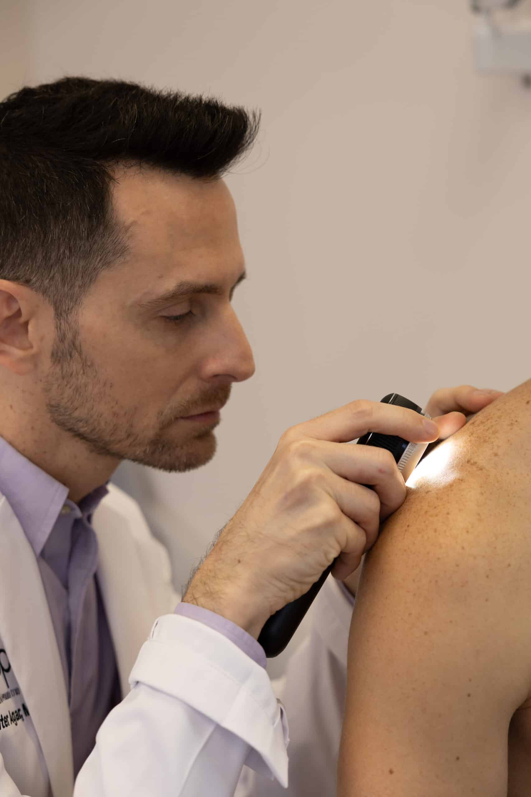
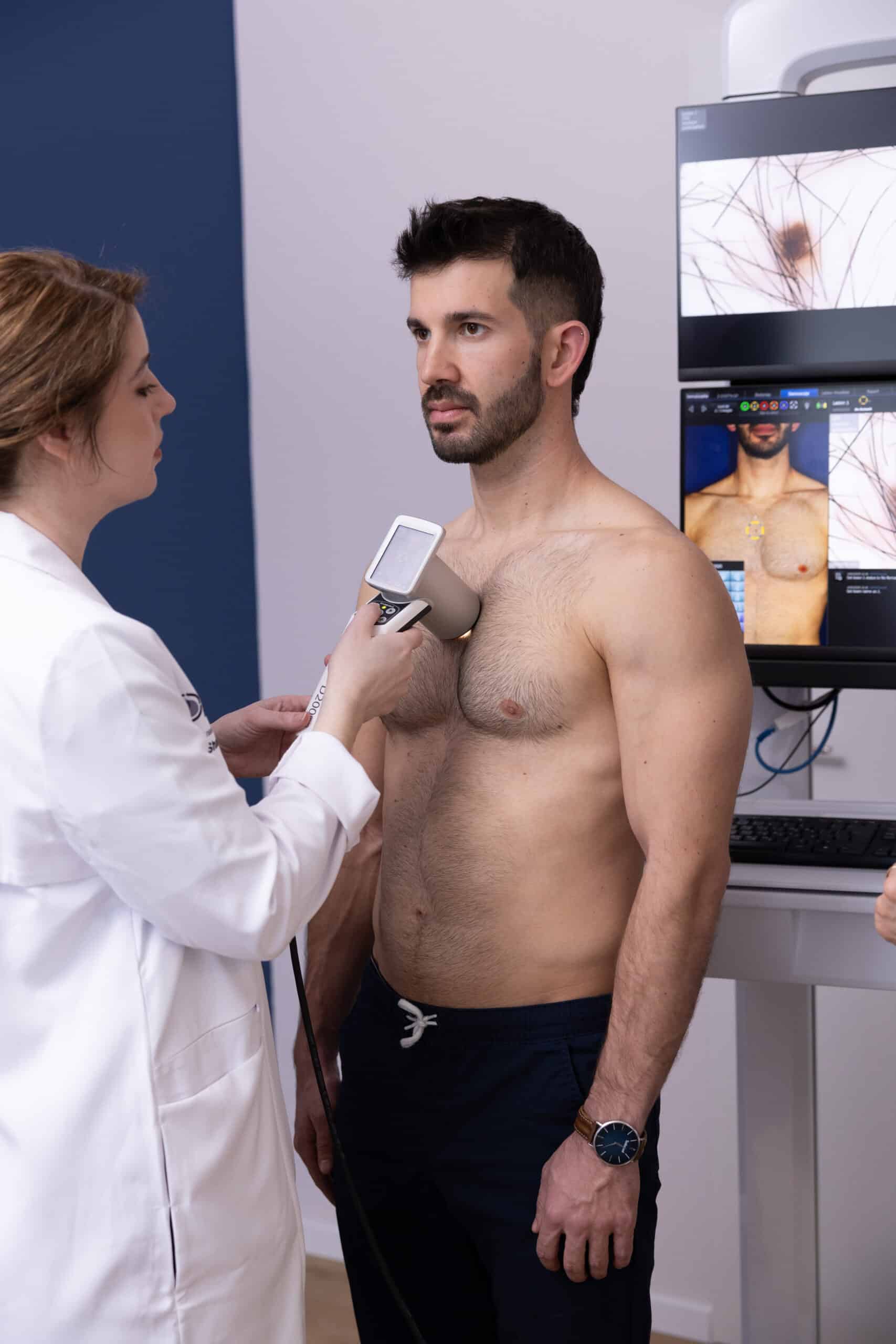
These tumors typically appear on sun-exposed areas (face, neck, arms) as raised, shiny lesions that range from skin-colored to pink and may occasionally have darker pigmentation. They are usually painless and non-itchy but may bleed easily or spontaneously, later forming a scab. Many patients are diagnosed with BCC after investigating a non-healing wound. Early diagnosis greatly increases the chance for cure using simple treatments and reduces the risk of recurrence.
BCC usually develops slowly, allowing for relatively easy treatment and full recovery in most cases. If left untreated, however, the tumor can grow in width and depth, invade local tissues, cause chronic wounds that are prone to infection, and lead to significant disfigurement. BCC rarely spreads to distant organs, but in rare aggressive cases, it can. Thus, while BCC generally affects quality of life more than life expectancy, early treatment is essential to prevent complications and recurrence.
Knowing your risk level can help with prevention and early detection. Main risk factors include:
• Prolonged UV exposure (sun or tanning beds)
• Personal history of skin cancer (BCC, SCC, or melanoma)
• Fair skin
• Age over 50
• Male gender
• Past exposure to ionizing radiation (e.g., childhood treatment for tinea capitis)
• Genetic syndromes (e.g., Gorlin syndrome, Brooke-Spiegler syndrome)
• Immunosuppression due to disease or medications (e.g., autoimmune conditions, transplant patients)
Early detection combined with advanced drug therapies offers a very optimistic outlook for patients at risk for BCC. The treatment approach is tailored according to the disease stage, so accurate staging is crucial. Our expert team designs the optimal treatment plan in collaboration with the patient.
Staging determines whether the disease is early (surgically resectable), locally advanced (unresectable), or recurrent. This is critical to understanding the threat to quality of life and to selecting the appropriate treatment and follow-up. Staging is primarily clinical and only occasionally relies on imaging. Early disease is managed by a dermatologist or plastic surgeon. Advanced or recurrent cases are handled by an oncologist and surgeon (typically a plastic surgeon).
• Early disease: Small BCC with minimal invasion, low risk of disfigurement. Treatment may be surgical or non-surgical.
• Locally advanced disease: Large BCC that invades surrounding tissue, causes disfigurement, and is not easily removed without major cosmetic or functional consequences. Treatment is systemic and/or radiation-based.
• Recurrent disease: BCC that has returned after previous treatment. Treatment depends on tumor size, location, prior therapies, and overall patient condition. A multidisciplinary team determines the approach.
• Metastatic BCC: Extremely rare. Cancer spreads to lymph nodes or distant organs. Diagnosis is confirmed by imaging (CT) and biopsy. Treatment is usually systemic (drug-based).
Thanks to the visibility of skin lesions, early detection is possible using the naked eye or optical tools. Early treatment improves survival and saves lives. Early-stage BCC is treated either surgically or with local, non-surgical therapies:
• Surgical excision: Standard excision with clear margins or Mohs micrographic surgery. Early removal ensures small defects and optimal aesthetic outcomes.
• Non-surgical therapies: At our center, we offer various treatment options for BCC based on size, location, and risk level. Each case is reviewed individually. Treatment decisions are made jointly by the dermatologist and oncologist with patient input. The earlier the diagnosis, the simpler the treatment.
Examples include local excision, topical medications, cryotherapy (liquid nitrogen), electrosurgery, and photodynamic therapy. Non-surgical options can be highly effective, with cure rates of ~80%, complete tumor regression, and no scarring in some cases.
Locally advanced, unresectable, or recurrent BCC is treated with biological therapies or immunotherapy. Radiation therapy may also be used. The oncologist determines the most appropriate approach.
In over 90% of BCC cases, mutations in the Hedgehog signaling pathway drive tumor growth. Oral medications (e.g., Erivedge, Odomzo) can effectively block this pathway, often leading to rapid and significant tumor regression in advanced or widespread disease. While similarly effective, each drug has a distinct side effect profile. The oncologist determines the best option and duration of treatment.
If targeted therapy fails, immunotherapy may be used to harness the patient’s immune system. BCC evades immune detection by exploiting natural checkpoints. Immunotherapy blocks these (e.g., PD-1) to help the immune system recognize and attack the tumor. Libtayo is the approved drug in this setting, administered via IV every few weeks. These treatments can lead to prolonged tumor regression. The oncologist tailors and monitors treatment.
הטיפולים אפקטיביים ומביאים לעיתים קרובות לנסיגת מחלה ממושכת. התאמת הטיפול והליווי נעשים ע”י האונקולוג בהתאם למאפייני המחלה והמטופל.
Patients with BCC face two main risks:
• New tumors: Those with BCC or other skin cancers are at high risk of developing additional BCCs due to genetic or environmental factors. Early detection by a dermatologist using optical tools is critical.
• Recurrence: Larger or more aggressive tumors, especially in immunosuppressed patients, are more likely to recur. Most recurrences are local. Follow-up is coordinated by the oncologist and dermatologist.
Given these risks, a multidisciplinary and coordinated follow-up is essential to maximize patient safety.
SCC usually appears as scaly patches on the skin that do not heal. These tumors may also bleed easily, itch, or be sensitive to touch, although most are not painful. SCC is often diagnosed when evaluating a crusted, ulcerated lesion. Many patients develop SCC in areas affected by actinic keratosis, a precancerous lesion that typically appears red, scaly, and raised. Early diagnosis significantly improves the chances of successful treatment and reduces the risk of recurrence.
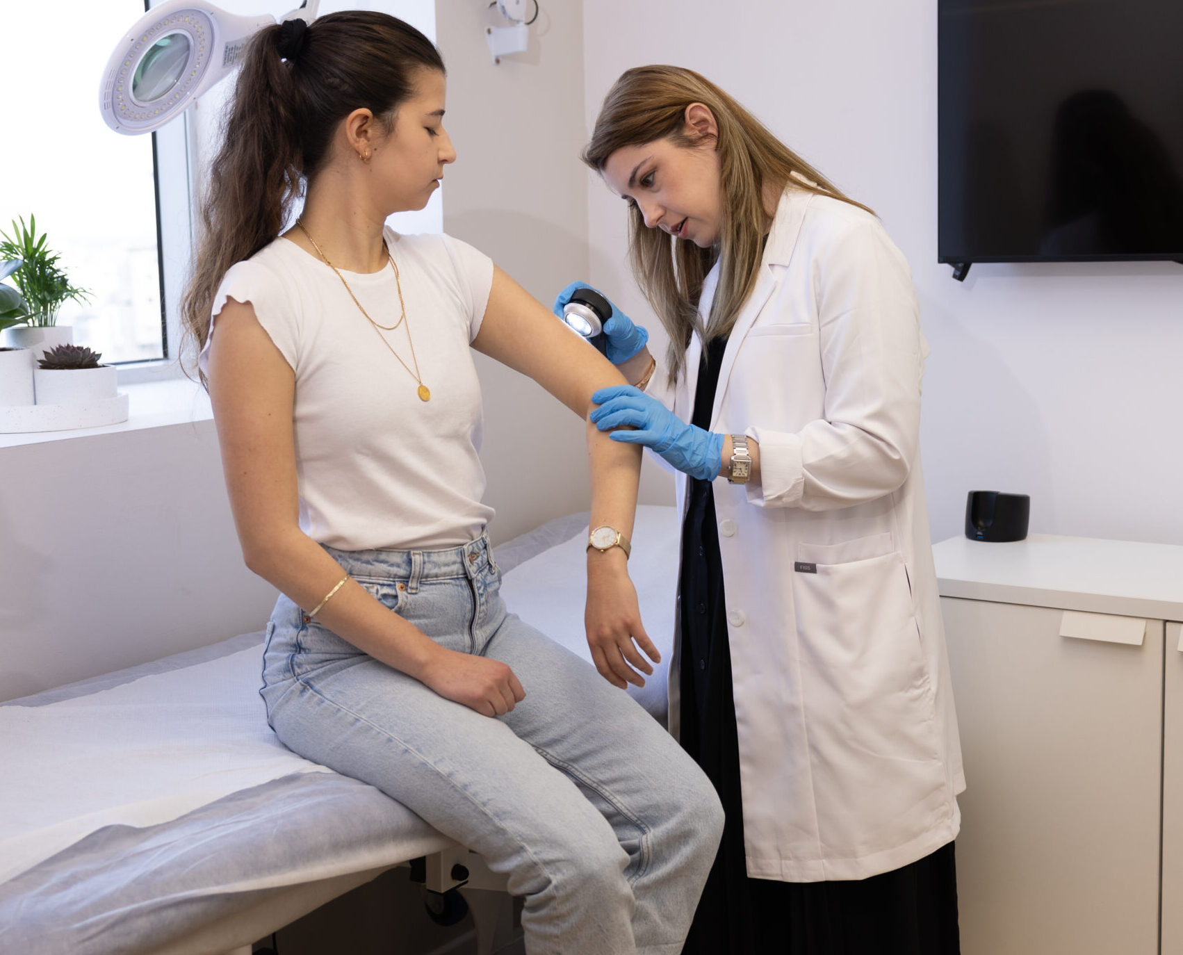
SCC typically grows slowly, and with timely treatment, complete cure is usually achievable. If left untreated, however, the tumor can spread both in depth and width, invade surrounding tissues, and metastasize to distant organs. In aggressive cases, this process can begin early. SCC can therefore threaten not only quality of life but also life expectancy. Early intervention is critical to reduce future risk.
Understanding your risk profile helps with prevention and early detection. SCC risk factors include:
• Prolonged UV exposure (sunlight or tanning beds)
• Personal history of skin cancer (BCC, SCC, or melanoma)
• Fair skin
• Immunosuppression due to disease or immunosuppressive medications (e.g., autoimmune disease or organ transplantation)
• Exposure to certain chemicals, especially occupationally
• Chronic skin inflammation (e.g., from burns, inflammatory skin diseases, or chronic wounds)
Early diagnosis and advanced therapies offer a positive outlook for SCC patients. Treatment is based on the disease stage, which must be carefully determined. Our expert team develops a tailored plan in full cooperation with the patient.
Staging determines whether the SCC is early-stage (surgically resectable), locally advanced (unresectable), metastatic, or recurrent. This classification is critical to planning treatment and follow-up and is typically based on clinical evaluation, occasionally supported by imaging. Early-stage disease is usually managed by a dermatologist, plastic surgeon, or oncologist. Advanced cases require a multidisciplinary team.
• Early-stage disease: Small SCC with minimal invasion, low risk of disfigurement or spread. Treated surgically or non-surgically. Radiation may be added postoperatively if the tumor has aggressive features or high recurrence risk.
• Locally advanced disease: Large SCCs that significantly invade surrounding tissues, often causing disfigurement. Surgery may result in major defects. Treatment is typically systemic and/or radiation-based.
• Recurrent disease: SCC that reappears after prior treatment. Treatment depends on tumor size, location, previous therapies, and patient condition.
• Metastatic SCC: SCC that has spread to lymph nodes and/or distant organs. Diagnosis is confirmed by imaging (e.g., CT scan) and biopsy. Treatment is usually systemic (drug-based).
Because SCC is visible on the skin, it can be detected early using the naked eye or optical tools. Early diagnosis directly improves survival. Early-stage SCC is typically managed with surgery or local non-surgical treatments:
• Surgical removal: Performed via standard excision or Mohs micrographic surgery. Early detection allows for smaller defects and better cosmetic outcomes.
• Non-surgical alternatives: SCC can be treated with various methods depending on size, location, and aggressiveness. Our center offers a full range of non-surgical options. Each case is evaluated individually, and treatment is selected jointly by our dermatology and oncology team with patient input. The earlier the diagnosis, the simpler the treatment.
Options include local excision, topical treatments, cryotherapy, electrosurgery, and photodynamic therapy. In selected cases, these methods have shown excellent success rates (~80%), complete lesion resolution, and no scarring.
Locally advanced, unresectable, or recurrent SCC is treated with advanced therapies such as immunotherapy. Radiation therapy may also be added. The oncologist will design the most appropriate treatment plan.
Immunotherapy is the first-line treatment for advanced SCC. It harnesses the patient’s immune system to recognize and destroy cancer cells. SCC evades immunity by exploiting internal regulatory mechanisms. Immunotherapy targets the PD-1 checkpoint and is administered intravenously every few weeks. Approved drugs include Libtayo and Keytruda, both highly effective. In many cases, they lead to significant long-term tumor regression. The oncologist will personalize the plan and monitor progress. If immunotherapy fails, chemotherapy combined with targeted biological therapy is considered.
Patients with SCC face dual risks:
• New tumors: Patients who have had SCC or other skin cancers are at higher risk of developing additional skin tumors due to genetic or environmental factors. Early detection by a dermatologist using optical tools is vital.
• Recurrence: Large or aggressive tumors, particularly in immunosuppressed patients, have higher recurrence risks. Most recurrences are local, but some may metastasize. Follow-up is jointly coordinated by the oncologist and dermatologist.
Multidisciplinary and coordinated follow-up is essential to ensure the highest level of patient care.
Merkel Cell Carcinoma (MCC) is a rare skin cancer that typically presents as a rapidly growing lesion over several weeks. It often appears purplish or reddish in color. MCC is a highly aggressive tumor with a high likelihood of spreading to lymph nodes or distant organs. If a new skin lesion appears and grows quickly, it is important to consult a dermatologist without delay. MCC is more common in older adults, and immunosuppression is a major risk factor. Treatment usually involves surgery and radiation. If the disease has spread to distant sites, immunotherapy is required.
On.Top is a specialized medical center dedicated exclusively to the diagnosis and treatment of melanoma and skin cancers.
Led by Prof. Gal Markel and Dr. Ofer Reiter Agar, two of Israel’s leading experts in dermatology and oncology, On.Top combines international clinical and academic excellence. It is one of the only centers in Israel that integrates multidisciplinary medical expertise under one roof — from initial diagnosis to oncologic follow-up — offering comprehensive, personalized care for high-risk patients.