דף הבית » Computerized Mole Mapping for Skin Cancer and Melanoma Diagnosis

At On.Top, you can be examined by a board-certified dermatologist using the most advanced technology for early diagnosis and detection. The total body photography system provides comprehensive digital mapping of the entire skin surface, allowing for in-depth evaluation of moles and lesions to detect suspicious findings. This significantly improves the chances of early detection of melanoma and other skin cancers, especially for individuals at increased risk.
Technological advancement and artificial intelligence are now at the service of medicine, enabling more precise and life-saving diagnoses than ever before.
In digital mole mapping, the entire body is scanned using a high-resolution camera that captures detailed images of the skin. This allows the dermatologist to monitor moles, lesions, and other skin changes over time. The system creates a complete digital map of the body, including close-up images of any suspicious areas. All images are stored securely and can be easily reviewed and compared in future visits. This enables accurate monitoring of changes over time without relying on memory alone—typically every few months or annually, according to the dermatologist’s recommendation.
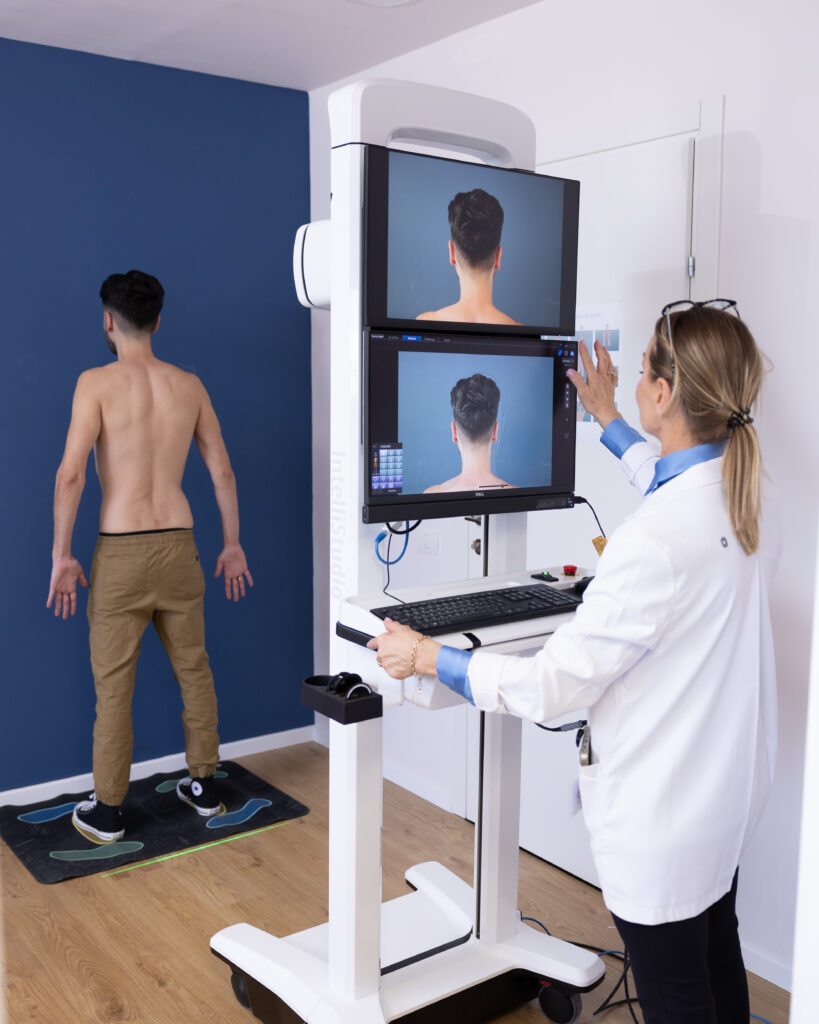
The system automatically detects changes using AI technology, as well as through manual review by a dermatology expert. It highlights changes in size, color, borders, or texture, allowing differentiation between stable benign lesions and new or evolving lesions that may indicate skin cancer.
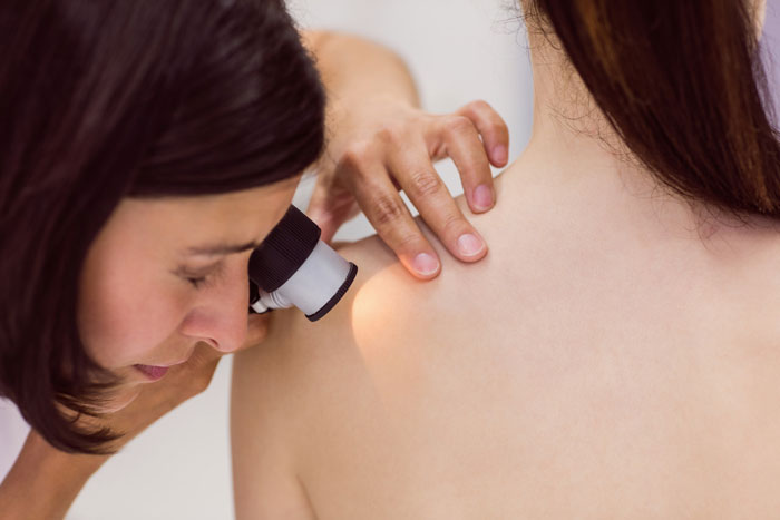
If a lesion appears unclear, it is further evaluated with digital dermoscopy—high-resolution imaging using a dermatoscope at intervals of 3–6 months. Comparing these images over time helps detect even the most subtle changes that may raise concern for malignancy, enabling very early cancer detection.
This precise follow-up also helps avoid unnecessary biopsies for lesions that remain unchanged throughout imaging follow-up.
The procedure is completely painless, involves no radiation, and has no side effects.
נגע עורי יוצא דופן, שנראה שונה משאר נגעי העור
גילוי נגע שלא היה קודם
נגע המשנה את צורתו או גודלו, או שלפתע מתחיל לדמם
לבעלי סיכון מוגבר למלנומה ולסרטן העור מומלץ לבצע אבחון מוקדם ובדיקת סקר אחת לחצי שנה עד שנה. חייבים לזכור כי מטופלים המאובחנים עם סרטני עור, ובמיוחד מלנומה בשלב מוקדם, הם בעלי אחוזי שרידות גבוהים מאוד ולרוב לסרטן אין כל השפעה על אורח או תוחלת החיים. לעומת זאת, מטופלים המאובחנים עם סרטני עור ומלנומה בשלב מתקדם הינם בעלי אחוזי שרידות נמוכים יותר ונזקקים לניתוחים מורכבים ולטיפולים תרופתיים.
בנוסף, קיימת הסכמה רחבה של הארגונים המדעיים לגבי הצורך של מטופלים עם היסטוריה של מלנומה להישאר במעקב לכל חייהם אצל רופא עור ובמקרים מסוימים גם רופא אונקולוג. תדירות המעקב המומלצת משתנה, בין היתר, כתלות במשך הזמן שחלף מאז האבחנה של המלנומה. התדירות נעה בין אחת לשלושה חודשים עד אחת לשנה.
קיימים גורמי סיכון נמוכים יותר כמו עור בהיר, שיער ג’ינג’י, עיניים כחולות או ירוקות, והיסטוריה של כוויות שמש או מצבים של מערכת חיסונית בדיכוי (כתוצאה מסרטן, מחלות אוטואימוניות או נטילת תרופות המדכאות את מערכת החיסון כגון במטופלים מושתלי איברים) בכל מקרה, ההמלצה היא לשמור על ערנות גבוהה גם אם לא נכללים בקבוצת הסיכון, ולשים לב לכל הופעה או שינוי של נגע חשוד- נקודת חן, שומה או כתם.





At our center, you’ll find a dedicated team of experts — experienced medical professionals focused on the diagnosis and treatment of melanoma and skin cancer, along with an administrative team to support you with all logistics and coordination.
To ensure the most accurate and comprehensive evaluation, please follow these guidelines before your appointment:
Not at all. It is a completely non-invasive and painless process—essentially a full-body ‘selfie’ without any radiation or side effects.
The recommended frequency for repeat total body mapping depends on your individual risk factors and the dermatologist’s clinical assessment.