

Nail Lesion Diagnosis

Evaluation and Management of Melanonychia

At our center, we offer diagnosis and treatment for changes in the nails that may indicate malignancy of the nail unit. We are here for the initial evaluation of any lesion, discoloration (brown or red), or subungual thickening, and provide follow-up treatment accordingly.

Melanonychia refers to the appearance of a brown or black band on the fingernails or toenails. It can result from various causes, including trauma, infections, systemic diseases, benign conditions, or malignancy. Consulting a dermatologist is the first step in diagnosing and ruling out serious conditions such as melanoma. At our center, we conduct comprehensive evaluations to ensure accurate diagnosis and proper management.
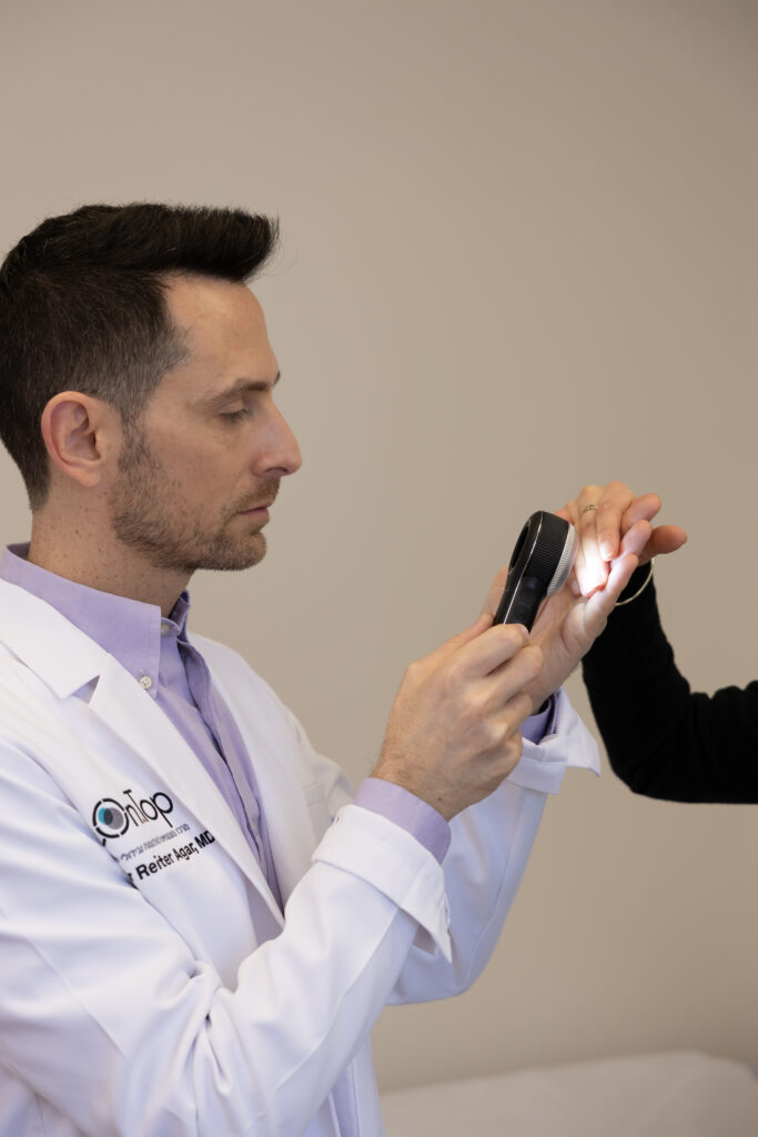
Our Diagnostic Approach Includes:

  1. Medical History
    The dermatologist gathers information on overall health, history of nail trauma or infections, medications, and family history of nail disorders or melanoma. This helps identify potential causes and risk factors.
  2. Physical Examination
    A thorough examination of the affected nails is performed to assess the characteristics of the melanonychia. This includes evaluation of the pattern, width, and location of pigmentation, as well as inspection of the surrounding tissues for signs of inflammation or infection.
  3. Dermoscopy
    Nail dermoscopy (onychoscopy) is a non-invasive technique using a specialized device to examine the nail plate and surrounding structures. Dermoscopy helps differentiate between benign and malignant causes of melanonychia based on identifiable features.
  4. Digital Dermoscopic Monitoring
    In borderline cases, high-resolution dermoscopic images may be taken over time to track the lesion’s progression. Monitoring enables detection of changes suggestive of malignancy, helping to avoid unnecessary invasive procedures.
  5. Biopsy
    If the diagnosis remains uncertain or there is suspicion of malignancy, a biopsy may be recommended. This involves taking a small tissue sample from the nail matrix, the growth area of the nail, for histopathological evaluation. Biopsy helps confirm the diagnosis and detect malignant cells if present.

Treatment Options

Treatment of melanonychia depends on the underlying cause and diagnostic findings. At our center, options are tailored to the individual and include:

  • Observation Only:
    If melanonychia is attributed to benign causes such as trauma or medication, monitoring may be sufficient. We observe for any changes in size, shape, or color to ensure the condition remains stable.
  • Treatment of Underlying Conditions:
    When melanonychia is secondary to infection, medication, or systemic disease, managing the underlying condition may resolve the discoloration. This may involve antifungal or antibacterial treatment, correction of nutritional deficiencies, or management of systemic illness.
  • Surgery:
    In cases of suspected melanoma or other malignant lesions, surgical intervention may be required. This typically includes excision of the affected area of the nail matrix or nail bed, along with a margin of healthy tissue.

We are committed to providing accurate diagnoses and effective, personalized treatment plans for every patient.

In Summary

If you notice any changes in the color or shape of your nails, we recommend scheduling an appointment promptly with our expert dermatologists for a thorough evaluation.


 

Recognized by most insurance companies
הפניקס
הראל
כלל ביטוח
מנורה מבטחים
קבוצת מגדל

You’re welcome to book an appointment – simply fill in your details and send.

Frequently Asked Questions

Frequently Asked Questions

At our center, you’ll find a team of experienced specialists—medical staff focused on diagnosing and treating melanoma and skin tumors, alongside a service team that handles all administrative matters.

Is a nail examination painful?

Typically, an initial nail examination is not painful, as it mainly involves an external, non-invasive inspection using visual observation or a dermoscope.

If there is suspicion of a fungal or bacterial infection, the physician may recommend a fungal test, which includes scraping the surface of the nail to collect a sample. This procedure might cause slight discomfort but should not be painful

It is recommended to avoid manicures or pedicures for at least a week before the examination. This allows the nail to return to its natural appearance and prevents interference from external substances.

The physician needs to examine the nail in its natural state to identify features such as pigmentation bands, thickness, or changes in the nail plate. Nail polish, gel, or deep filing can conceal important changes in nail structure, color, or texture that are critical for diagnosis.
Chemical products used in manicures or pedicures may also affect laboratory test results. Additionally, aggressive treatments can cause minor injuries, inflammation, or infections, which may complicate the diagnostic process.

It is best to keep your nails clean and dry prior to the examination.

Not every dark stripe on the nail is a sign of melanoma, but it’s important to take it seriously and investigate the cause. Dark stripes can result from a variety of reasons—some benign and others that require medical attention.

Warning signs to look out for include:

  • A new dark stripe, especially in people with fair skin
  • A stripe that is widening or spreading over time
  • Irregular color or texture within the stripe
  • Changes in the skin around the nail
  • Bleeding, pain, or nail breakage
  • Rapid changes such as a sudden appearance of a stripe, evolution in its appearance, or accompanying symptoms like inflammation or discomfort

In any case, it’s essential to monitor for unusual features or changes. Early detection is crucial—even when it comes to nails.

Complementary Services

Mole Mapping

Full-body digital dermoscopy imaging

Skin Biopsy

Tissue sampling for further analysis

Patient Testimonials for the Specialists Center

×

WhatsApp Chat

השאירו פרטים ונחזור אליכם בקרוב
Skip to content