

The first step in diagnosis and treatment starts here — with a skin check that should be part of your routine health maintenance. Just like blood tests, dental cleanings, or breast cancer screening, your skin also requires regular evaluation, especially when new moles, spots, or suspicious lesions appear.
Accurate diagnosis and early detection are key to life-saving treatment in cases of melanoma or other skin cancers.
The initial evaluation is relatively simple, non-invasive, and involves no side effects or radiation exposure. It is performed by our dermatologists, who specialize in early detection and lesion assessment.
Suspicious skin lesions may be pigmented (brown to black) or non-pigmented (skin-colored to pink). However, pigmentation alone does not determine the nature of a lesion — both benign and malignant lesions can be pigmented or non-pigmented.
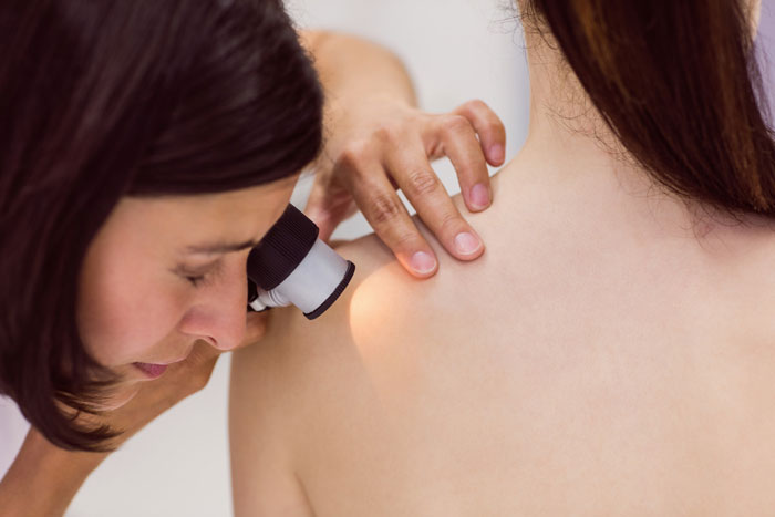
The exam includes a full-body skin check with the naked eye, along with a thorough dermoscopic evaluation.
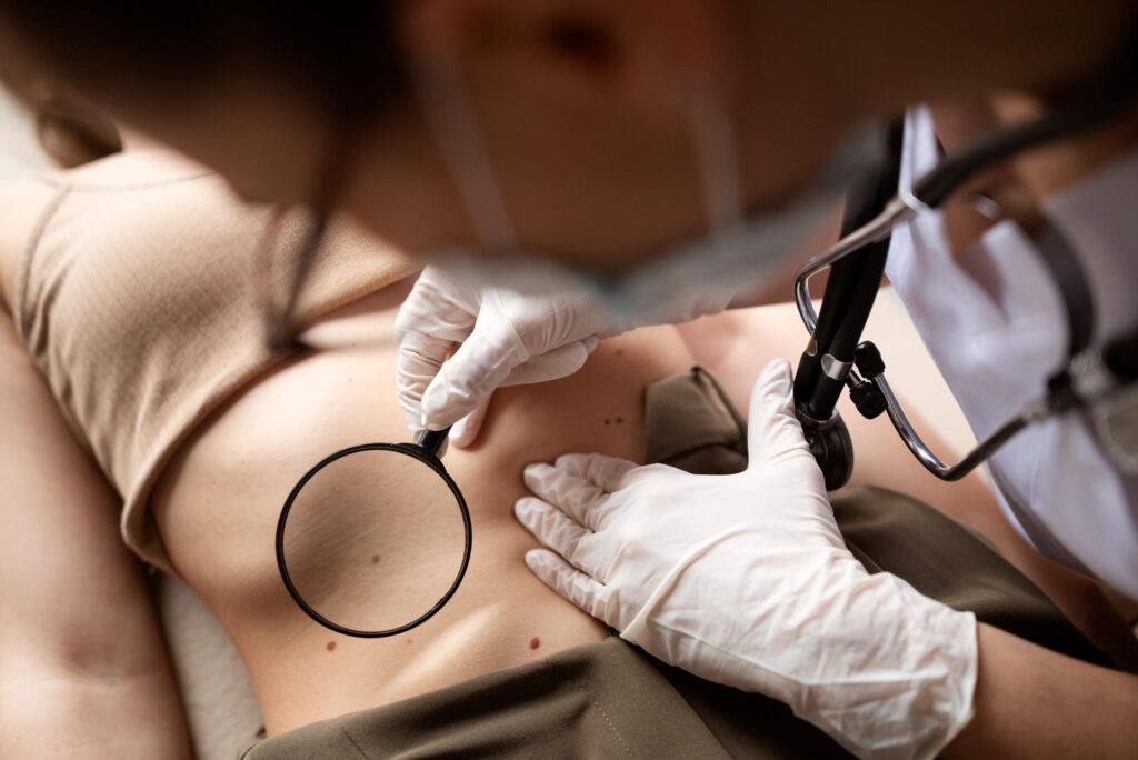
Often called the “stethoscope of dermatologists,” the dermatoscope allows for far more accurate diagnosis of skin cancer than visual inspection alone.
It consists of a magnifying system, polarized light source, and a filter. The way the light is transmitted into the skin and reflected back through the filter and lens allows the physician to visualize structures within the upper layers of the skin not visible to the naked eye.
A dermatologist trained in dermoscopy can often determine whether a lesion is benign or requires further investigation, including a surgical biopsy.
We strongly encourage incorporating a skin check into your regular health checkups.
Because some lesions can be difficult to detect during self-examination, we recommend a yearly skin exam for individuals who are not in a high-risk group but have 20 or more moles/lesions.
A skin lesion that looks different from the rest
A spot that recently appeared and wasn’t there before
A mole or spot that changes shape, size, or begins to bleed
Skin cancer is one of the most common cancers worldwide, and virtually anyone can be affected. However, certain individuals are at significantly higher risk, including those with:
Additional risk factors include:
Even if you don’t fall into a high-risk group, it’s still essential to stay alert and consult a dermatologist about any new or changing spots, moles, or lesions.
There are additional, lower-level risk factors such as fair skin, red hair, blue or green eyes, and a history of sunburns, as well as conditions involving immune suppression (due to cancer, autoimmune diseases, or the use of immunosuppressive medications, such as in organ transplant patients).
In any case, the recommendation is to remain highly vigilant—even if you are not part of a recognized high-risk group—and to pay attention to any new or changing suspicious lesion, such as a mole, birthmark, or skin spot.





At our center, you will find a team of experts – experienced medical professionals specializing in the diagnosis and treatment of melanoma and skin cancers, along with a dedicated administrative support staff.
The initial screening does not require any special preparation. The examination is external and covers the entire skin surface—both visible and less easily noticeable areas. It is recommended to inform the doctor about any suspicious lesions or changes you may have noticed.
The examination is painless and does not cause any physical discomfort. It is a non-invasive, external assessment performed with quiet and non-intrusive equipment. The process is simple and is recommended as a routine check-up once a year.
The results of the initial screening are provided immediately at the end of the examination. Based on the findings, the physician will decide whether additional, more detailed evaluation is required. If there is suspicion of a lesion that needs further investigation, you will be updated right away regarding the next steps and how the evaluation will be carried out.